SOPHiA DDM™
for Radiomics

Unlock entirely novel insights from your radiology images
Generate new insights from your existing radiology workflows through AI-powered radiomics analytics with expert-driven visualization, segmentation, and extraction functionalities in one user-friendly interface.
Request a Demo
Home breadcrumb-arrow SOPHiA DDM for Radiomics

Explore new horizons in your radiomics research projects

The SOPHiA DDM™ Platform is compatible with any 3D imaging modality, equipment, and cancer type. 
Gain novel insights through image visualization and automated processing in a collaborative, multicenter research environment.
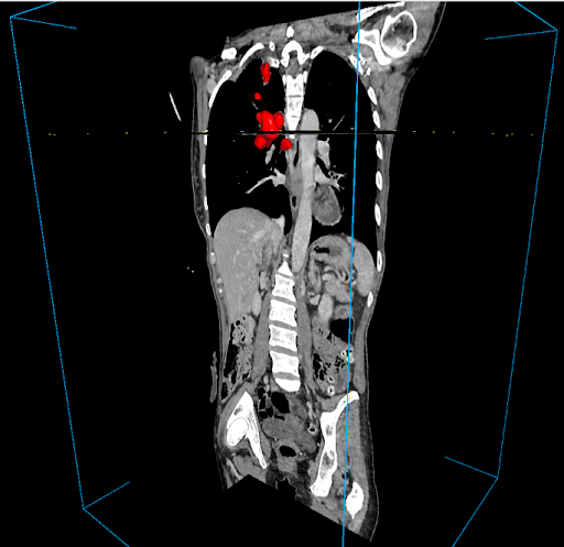
Streamlined 3D 
segmentation
Automatically or semi-automatically segment anatomical areas of interest in a matter of seconds.
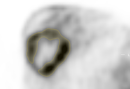
Reliable feature
extraction
Leverage validated methods for automated extraction of radiomic features.
Collaborative
environment
Empower multicenter studies with a multimodal research database accessible by anyone, anywhere.
Streamlined 3D
segmentation
Automatically or semi-automatically segment anatomical areas of interest in a matter of seconds.
Reliable automatic
feature extraction
Leverage validated methods for automated extraction of radiomic features.
Collaborative
environment
Participate in multicenter studies across our global network, utilizing our multimodal research database.

Democratizing
data-driven medicine

Improve accuracy

Utilize robust AI-powered algorithms to derive deep insights from complex data.

Expedite turnaround times

Utilize robust AI-powered algorithms to derive deep insights from complex data.

Accelerate adoption of precision medicine

Utilize robust AI-powered algorithms to derive deep insights from complex data.

Optimize resources

Utilize robust AI-powered algorithms to derive deep insights from complex data.

Optimize resources

Utilize robust AI-powered algorithms to derive deep insights from complex data.

Easily navigate the path from images to insights

Import 3D images (e.g., CT, MRI, and PET scans) to a project-specific database and research environment.
Visualize images in 3D, from multiple angles at the same time with the user-friendly interface.
Automatically or semi-automatically segment anatomical areas of interest using our proprietary algorithms.
Utilize robust AI-powered algorithms to derive deep insights from complex data.
Confidently extract radiomic features from segments and store in the research environment for further analysis.
Analyze radiomic features and add data to multimodal cohorts and longitudinal analyses.

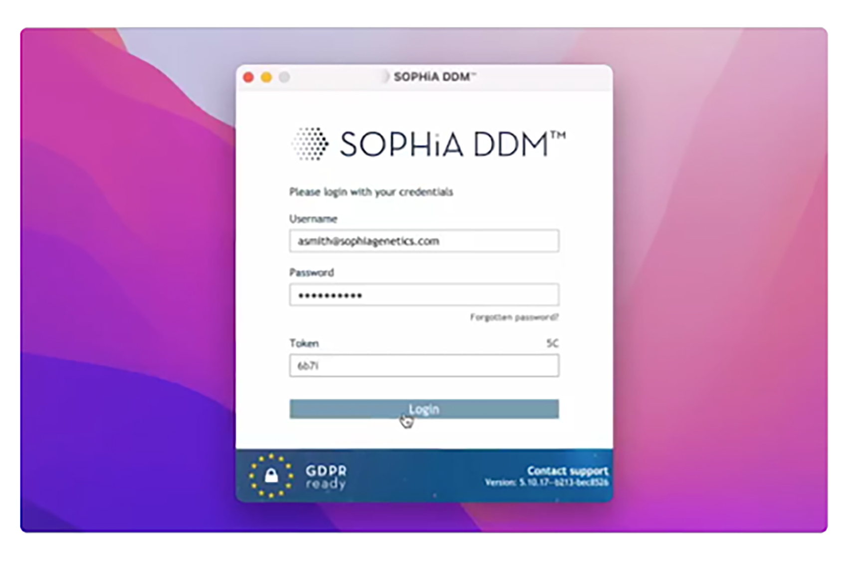
Securely log in to our cloud-based platform with 2-step verification. Quickly upload FASTQ files and select from performance pipelines tailored to sample type, chemistry, enrichment kit, and sequencer.

Reaching new heights in precision medicine

Metastatic non-small cell lung cancer
A machine learning model was developed for the TRIDENT post hoc analysis of AstraZeneca’s POSEIDON trial to identify multimodal signatures predicting which mNSCLC patients would benefit most from adding tremelimumab to durvalumab + chemotherapy. Radiomics was a key contributor to the model’s predictive power.
Read Abstract
Triple-negative breast cancer
A machine learning algorithm applied to baseline multimodal data of a retrospective cohort was developed to predict complete pathological response to neoadjuvant chemotherapy in TNBC. Radiomics was one of the key contributors to the predictive power of the model.
Read Abstract
Solid renal tumors
In the UroCCR-75 study, a machine-learning model was developed to differentiate benign from malignant solid renal tumors. The model was based on clinical, radiologic, and radiomics features from pre-operative multiphasic contrast-enhanced CT scans from a national kidney cancer database.
Read Abstract
Meningioma
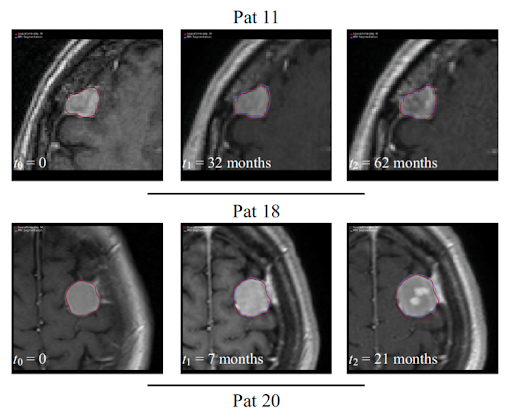
Spatial mechanistic modeling was used to predict growth of benign asymptomatic meningiomas aiding decisions on whether to extend follow-up or treat the tumor. This approach was able to predict changes in tumor volume and shape after only two MRI examinations, outperforming naïve linear regression.
Read Abstract
Colorectal liver metastases
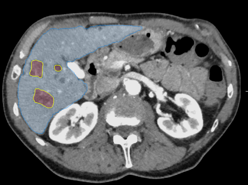
A post hoc evaluation of the TRIBE2 study on liver-limited metastatic colorectal cancer found that radiomics may be a valid tool for predicting prognosis in patients receiving first-line treatment, and for stratification according to risk of relapse after curative resection.
Read Abstract

Lorem ipsum dolor aset

Lorem ipsum dolor sit amet, consectetur adipiscing elit. Mauris semper dolor ut velit tristique, quis dignissim dui ullamcorper. Proin viverra leo eu ullamcorper consequat. Sed sit amet interdum est. Integer porttitor dui a augue tincidunt, ac pellentesque orci lacinia.

Easily navigate complex analysis

SOPHiA DDM™ for Blood Cancers simplifies analysis of genomic drivers in hematological cancers using the trusted analytical performance and advanced features of the SOPHiA DDM™ platform.
Streamline raw genomic data analysis to precisely detect, annotate, and prioritize even the most complex blood cancer variants.

Heading for References lorem ipsum

SOPHiA GENETICS products are for Research Use Only and not for use in diagnostic procedures unless specified otherwise.

SOPHiA DDM™ Dx Hereditary Cancer Solution, SOPHiA DDM™ Dx RNAtarget Oncology Solution and SOPHiA DDM™ Dx Homologous Recombination Deficiency Solution are available as CE-IVD products for In Vitro Diagnostic Use in the European Economic Area (EEA), the United Kingdom and Switzerland. SOPHiA DDM™ Dx Myeloid Solution and SOPHiA DDM™ Dx Solid Tumor Solution are available as CE-IVD products for In Vitro Diagnostic Use in the EEA, the United Kingdom, Switzerland, and Israel. Information about products that may or may not be available in different countries and if applicable, may or may not have received approval or market clearance by a governmental regulatory body for different indications for use. Please contact us at support@sophiagenetics.com to obtain the appropriate product information for your country of residence.

All third-party trademarks listed by SOPHiA GENETICS remain the property of their respective owners. Unless specifically identified as such, SOPHiA GENETICS’ use of third-party trademarks does not indicate any relationship, sponsorship, or endorsement between SOPHiA GENETICS and the owners of these trademarks. Any references by SOPHiA GENETICS to third-party trademarks is to identify the corresponding third-party goods and/or services and shall be considered nominative fair use under the trademark law.

SOPHiA DDM™ Overview
Unlocking Insights, Transforming Healthcare
Learn About SOPHiA DDM™ 
SOPHiA DDM™ for Genomics

Oncology 

Rare and Inherited Disorders

Add-On Modules

SOPHiA DDM™ for Radiomics
Unlock entirely novel insights from your radiology images
Learn About SOPHiA DDM™ for Radiomics 
SOPHiA DDM™ for Multimodal
Explore new frontiers in biology and disease through novel insights
Learn About SOPHiA DDM™ for Multimodal
Professional Services
Accelerate breakthroughs with our tailored enablement services
Learn About our Professional Services